


Biology For Class X - Chapter No. 3 - Coordination And Control - Long Question Answers
GO TO INDEX
CHAPTER 3: COORDINATION AND CONTROL
C. Extensive Response Questions And Notes
Q.1: Discuss Nervous system of man.
Ans: Nervous System of Man:
It controls all functions of the body.
Human nervous system like other vertebrates is “centralized-type nervous system” (CNS) and the most advanced, highly developed. It is the most complicated type. Stimuli from various organs of the body are sent simultaneously its control center or central nervous system where they are integrated, analyzed and processed to develop command in the form of response.
It consists of two major divisions:
- Central Nervous System (CNS)
- Peripheral Nervous System (PNS)
1. Central Nervous System (CNS):
It is the major command and control center to which stimuli are reported and decisions are made and conveyed to effector organs. The Central nervous system(CNS) consists of brain and spinal cord and also consists of up to 100 billion inter neurons.
Components of Central Nervous System:
The central nervous system consists of
- Brain and
- Spinal Cord
2. Peripheral Nervous System (PNS):
The cables or nerves which arise from the central nervous system (i.e brain and spinal cord), are spread in various part of the body and transmits the signals between CNS and body parts make a nervous system called Peripheral Nervous System. Each nerve consists of bundles of axons of both sensory and motor neurons.
Types of Peripheral Nervous System:
The peripheral nervous system is divided into two types according to their functions.
- Somatic Nervous System:
The peripheral nervous system which controls all the voluntary activities of the body such as contraction of skeletal muscles and movement of joint is called Somatic Nervous System. - Autonomic Nervous System:
The peripheral nervous system which controls involuntary activities of the body such as smooth muscles, glands, muscles of heart, and other internal organs (like for digestion, breathing, etc.) is called Autonomic Nervous System. These involuntary activities or functions are vital for maintaining life processes.

Ans: HUMAN BRAIN
The most important part of Central Nervous System develops from dorsal, hollow nerve cord well protected in the cranium of skull and composed of inter neurons and is the seat of our intelligence, learning and memory is called Brain. It is the major command and control center of our body.
Brain is protected in three ways:
- Cranium: Bones of the skull provide protection to the brain.
- Meninges: Beneath the cranium, the brain and spinal cord are wrapped in three protective membranes made up of tough connective tissues called meninges.
- Cerebrospinal Fluid (C5F): Between the layers of meninges, there is a plasma like fluid which bathes the neurons of brain and spinal cord is called Cerebrospinal Fluid (CSF). It also provides a cushion-like protection to the brain.

The brain consists of Following important parts
- Cerebrum
- Hippocampus
- Amygdala
- Thalamus
- Hypothalamus
- Mid brain
- Cerebellum
- Medulla oblongata
i. CEREBRUM:
Cerebrum is the largest part of the brain where all important decisions are made . It is divided into two halves called hemispheres.
-
Right cerebral hemisphere:
The right cerebral hemisphere regulates the left side of the body. -
Left cerebral hemisphere:
The left cerebral hemisphere regulates the right side of the body.
i. An outer grey matter or cerebral cortex and
ii. An inner white matter
ii. An inner white matter
- Cerebral Cortex OR Grey matter:
Cerebral cortex or grey matter is the outer part of cerebrum and is grayish in colour. It is largest and the most complex part of human brain. It is highly convoluted to occupy the greater number of inter neurons. - White Matter:
The inner part is white in colour and called the white matter. The white matter consists of cell processes which are hair like growths.
The cortex is associated with thoughts, plans, actions and determination.
Part of Cortex:
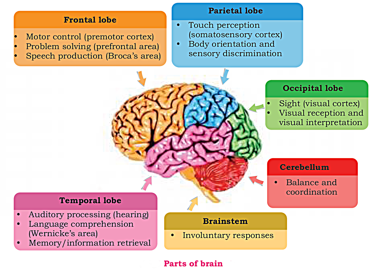
Functionally, It can be divided into four sections or lobes, viz.:
- Frontal lobe:
It is associated with thoughts, emotions, etc.
• Motor control (premotor cortex)
• Problem solving (prefrontal area)
• Speech production (Broca's area) - Parietal lobe:
It is associated with different sensations like pressure, temperature, language processing, etc.
• Touch perception (somatosensory cortex)
• Body orientation and sensory discrimination - Temporal lobe:
It is involved in hearing and speech.
• Auditory processing (hearing)
• Language comprehension (Wernicke's area)
• Memory/information retrieval - Occipital lobe:
It is associated with vision.
• Sight (visual cortex)
• Visual reception and visual interpretation
Function of Cerebrum:
- Cerebrum is concerned with seat of intelligence, memory, learning, reasoning and overall control of all voluntary actions.
- It involved in all conscious activities.
- It co-ordinates different senses together.
ii. THALAMUS:
Thalamus lies inside the brain just above hypothalamus.
Functions:
- It is also called the clearing house of sensory impulses.
- It receives stimuli from different parts of brain and relays them to the appropriate part of the cortex.
iii. HYPOTHALAMUS:
Hypothalamus is the part of limbic system which is called Thermostat of the body. It regulates life maintaining functions.
Functions:
- The hypothalamus is important in regulation of homeostasis of the body.
- It regulates pituitary gland.
- It also regulate body temperature, blood pressure, hunger, thirst, aggression, pleasure and pain.
iv. HIPPOCAMPUS:
Hippocampus is involved in long term memory.
v. AMYGDALA:
It is a deep seated small area.
Function:
- The amygdala produces sensation of pleasure, punishment or sexual arousal stimulation.
- It also involve in the feelings of fear.
vi. MID BRAIN:
In mammals mid brain is relatively very small. It consists of the optic lobes which are represented by four small bodies.
Functions:
- It receives sensory information like vision, olfactory (odour, smell) etc.
- It receives sensory information from spinal cord and sends them to the fore brain.
vii. CEREBELLUM:
The cerebellum lies dorsally behind the optic lobes. It is highly convoluted. It is large in mammals than other animal.
Functions:
• Balance and coordination
- The cerebellum plays an important part in controlling muscular co-ordination.
- It specially controls the precision in movement of the muscles for balance and maintains the position of the body in relation to gravity.
- Activities like writing, drawing, painting, dancing, crafting have become possible due to its elaborate structure in human.
viii. MEDULLA OBLONGATA:
Medulla oblongata lies on the top of spinal cord.
Functions:
- It is the control center for automatic activities like breathing, heart-beat, blood pressure, coughing, swallowing, hiccuping, digesting food, etc. Such activities are termed as Reflexes.
ix. PONS:
It lies on the ventral side of medulla oblongata.
Functions:
- It helps in controlling the facial muscles as well as helps in sleep and wakening.
- Pons regulates activities like muscular co-ordination, and breathing.
BRAINSTEM
Midbrain, pons, and medulla is collectively known as brain stem. It connects cerebrum to spinal cord.
Function:
• Involuntary responses
Q.3: Write a short note on spinal cord?
Ans: Spiral Cord:
A butterfly shaped, thick whitish nerve cord that lies below the medulla oblongata and extends down through the neural canal of vertebrate up to the hips is called Spinal Cord.
Cross Section of Spinal Cord:
In cross section, the spinal cord is differentiated into two areas.
- White Matter
- Gray Matter
Funtion:
- It acts as a mini control center for few reflexes.
- It also acts as express way for flow of information from brain to the different parts of the body and vice versa.
Q.4: What do you know about neurons.
Ans: Definition Of Neuron:
The special cells of nervous system which can generate and conduct electric current are called Neurons. They are the chief structural and functional unit of nervous system.
Structure of Neurons:
Neurons are different from each other according to size and shape but the structure of neuron consists of two parts:
- Soma or Cell Body
- Cell processes
Soma OR Cell Body:
- Each neuron has a cell body or soma comprised of plasma membrane containing nucleus and various organelle embedded in the cytoplasm.
-
Functions:
The cell body or soma is the main nutritional part of the cell necessary for growth of neuron.
Cell Processes:
They are hair like projections given out from soma. Cell processes are usually enclosed by a layer of fatty substance known as Myelin Sheath. Like an electric cable, this serve as insulating layer to ensure the uninterrupted transmission of nerve impulse.
Types Of Cell Processes:
They are of two types:
-
Dendrites:
From the soma, a large number of small threads like terminal branches are arises are called Dendrites.
Functions:
Dendrites receive stimuli and convey it to the soma. - Axon:
The unbranched, single elongated cytoplasm process which usually arises opposite to dendrites called Axon.
Functions:
Axon is specialized for conducting impulses to other neurons or some effector cells.
Function Of Neuron:
Each neuron is specialized to generate and conduct neuronal signal or nerve impulse.

Q.5: Define and explain reflex action with the help of an experiment of shin muscle of frog using battery?
Ans: Reflex Action:
The automatic involuntary responses which occur either due to internal or external stimuli are called Reflex Action.
OR
The automatic, pre-programmed responses regulated by CNS are termed as
reex actions. Some of them are directly regulated by brain while others
by spinal cord.Parts of Reflex Action:
Reflex action consists of
- Receptor: (Skin) receive stimuli.
- Sensory Neuron: It carries message from receptor to the Central Nervous System (CNS).
- Motor Neuron: It carries message from Central Nervous System (CNS) to the effector.
- Effector: (Muscle of gland) which perform action.
Path Of Reflex Action:
Reflex Arc:
The pathway of passage in impulse during a reflex action is called Reflex Arc.
Working Of reflex arc:
- A receptor in the skin detects a stimulus (the change in temperature).
- Sensory neurons send electrical impulses to relay neurons, which are located in the spinal cord.
- Motor neurons send electrical impulses to an effector.
- The effector produces a response (muscle contracts to move hand away).
Examples of Reflex Actions:
- If our hand touch any hot object, it is quickly withdrawn.
- Secretion of juices from the gland.
- Blinking of eyes.
- Contraction and expansion of lungs.
- Knee jerk
- Sneezing, hiccup, cough etc.

EXPERIMENT: Contraction of shin muscle (leg muscle) of Frog using battery.
Observation:
- Muscles contract when signals are provided through neurons by nervous system.
- In this experiment, a shin muscle is removed from dissected frog and is placed in methylene blue solution in a petri dish.
- We observe that when we artificially stimulate it by supplying power of a 12 volts D.C. battery, it contracts.
Apparatus:
- Dissecting box
- Frog
- Dissecting tray
- Petri-dish
- 12 volt D.C. charged battery
- Wires
Procedure:
- Dissect out a pithed frog to expose its shin muscles.
- Remove the shin muscle with sciatic nerve carefully and place in stretched condition in petri-dish.
- Connect the battery with wires and then touch the shin muscle at its beginning and end with wires.
- Now observe the muscle contraction.
- Repeat the experiment for three times.
Conclusion:
- Muscles contracts when provided signals through neurons by nervous system.

Q.6: Explain the structure of human eye in detail. Also define accommodation?
Ans: Human Eye:
The eye is an important and one of the most complex sense organ. It is photoreceptor and the organ of sight. It helps us in visualizing objects and also helps us in light, color and depth perception. It works on the principle of a simple camera, which collect light reflected from any object in front and help us see objects.
Structure of Human Eye:
A human eye is roughly 2.3 cm in diameter and is almost a spherical ball filled with some fluid. Each eye lies in a bony socket for protection. It consists of the following parts:
Sclera:
- The outer covering, a protective tough white layer called the sclera (white part of the eye).
- Working: It protects eyes.
Cornea:
- The front transparent part of the sclera is called cornea.
- Working: Light enters the eye through the cornea.
Aqueous humor:
- It is a small chamber behind cornea and is filled with watery fluid.
Iris:
- A dark muscular tissue and ring-like structure behind the cornea is known as the iris.
- The colour of the iris actually indicates the colour of the eye.
- Working: The iris also helps regulate or adjust exposure by adjusting the iris.
Pupil:
- A small opening in the iris is known as a pupil.
- Its size is controlled by the help of iris.
- Working: It controls the amount of light that enters the eye.
- Pupil reflex:
The pupil adjusts itself depending upon the intensity of light.
In case of bright light, it protects the retina by constricting itself so less amount of light falls on retina.
In dim light condition, the pupil dilates to allow more light to fall on retina.
Lens:
- Behind the pupil, there is a transparent crystalline convex structure called a lens.
- It diverts light to a layer of sensory cells or retina.
Ciliary body:
- Lens is suspended by a ring of circular muscles known as ciliary body.
-
Working:
By the action of ciliary muscles, the lens changes its shape to focus light on the retina.
It becomes thinner to focus distant objects and becomes thicker to focus nearby objects.
Vitreous humor:
- It is the main cavity of eye ball filled with clear gel.
- It is behind the lens
Retina:
- The innermost layer of eye is retina on which the image is formed by cornea and lens.
- It is a light-sensitive layer that consists of numerous nerve cells.
- Working: It converts images formed by the lens into electrical impulses. These electrical impulses are then transmitted to the brain through optic nerves.
Optic nerves:
Optic nerves has sensory cells and are of two types. These include cones and rods which upon stimulation convert light signals into nerve impulses and report them to the brain
- Cones:
Cones are the nerve cells that are more sensitive to bright light.
Working: They help in detailed central and colour vision. - Rods:
Rods are the optic nerve cells that are more sensitive to dim lights.
Working: They help in peripheral vision.

Accommodation:
It is an automatic process of altering focus to get sharper image of the near objects. The ciliary muscles contract allowing the elastic lens to become thicker and more convex. With age, the lens loses its elasticity and as a result, accommodation becomes increasingly difficult.

Q.7: Describe the defects of vision?
Ans: DEFECTS OF EYE:
Eye-Sight Defect:
- Short-Sightedness:
Short-sightedness or myopia refers to the difficulty in focusing distant object while the near objects are focused normally. - Long-Sightedness:
Long- sightedness or Hyperopia is the difficulty in focusing closer objects while distant vision is clear.
Treatment:
Both can be diagnosed and corrected by using appropriate glasses or contact lenses.

Colour blindness:
- Its a deficiency of vision in which one cannot distinguish certain colours such as blue and yellow or red and green.
- It is due to the defect in cones of retina.

Role of vitamin A with vision:
- Vitamin A is required for proper vision and needed for sensory cells of retina.
- It also helps the cornea to be well lubricated.
- Deficiency of vitamin A could lead to corneal ulcers and blindness.
Q.8: Describe Contribution of Ibn-al- Haitham and Ali- Ibn- Sina about the strucuture of eye and treatment of ophthalmic diseases?
Ans: Contribution of Ibn-al-Haitham about the strucuture of eye and treatment of ophthalmic diseases:
- Ibn-al-Haitham ,known as the "father of modern optics" due to great contribution in principles of optics and visual perceptions was a great Muslim mathematician, philosopher, astronomer and physicist of 11th century.
- He was the first person to consider vision as a result of bouncing back of light from an object and then enters our eyes.
- Books: His most important book on optics was "Kitab-ul-manazir".
Contribution of Ali-Ibn-Isa about the strucuture of eye and treatment of ophthalmic diseases:
- He was one of the most important Muslim ophthalmologists of medieval times.
- Book: In his famous book "Memorandum of the oculists" on ophthalmology, he described more than hundred different eye diseases and their treatment.
Q.9: Explain the structure and function of human ear in detail.
Ans: Human Ear:
The human ear, like that of other mammals, contains sense organs that serve two quite different functions:
- Hearing
- Balancing
Parts of Human Ear:
It consists of three parts:
- Outer ear
- Middle ear
- Inner ear
OUTER EAR:
The outer ear collects and transmits sound waves. It consists of:
- Pinna: The pinna is composed of folds of skin and cartilage. It leads into the ear canal which is closed at the inner end by tympanic membrane.
- Ear canal: Ear canal has hair and it produces wax to trap dust and small foreign bodies.
- Tympanic membrane or ear drum: The tympanic membrane which is also called the eardrum separates the outer ear from the middle ear. When sound waves reach the tympanic membrane they cause it to vibrate. The vibrations are then transferred to the tiny bones in the middle ear.
MIDDLE EAR:
The middle ear receives sound waves from air outside and transmits it into the fluid in the inner ear. It consists of:
-
A small cavity containing three small moveable bones
i) malleus
ii) incus and
iii) stapes - It is connected to inner nasal cavity through a small tube, called the Eustachian tube.
INNER EAR:
The inner ear transforms sound waves into nerve impulse. It consists of:
- Cochlea: It is the front membrane associated with hearing.
- Three semicircular canals: They are deep inside the skull bones and are associated with balance.

Role of ear in balance:
- The three semicircular canals are inter connected and lie at right angle to each other.
- They are connected to a swollen part called the vestibule.
- Semicircular canals are sensitive to gravity, position and movements of head. The changes are detected and reported to brain through nerve fibers.
- Semicircular canals and vestibule are involved in maintaining balance of the body in relation to gravity.

Q.10: What is endocrine system? Discuss the gland involved in regulation of blood glucose and how?
Ans: ENDOCRINE SYSTEM:
Endocrine system is an important means of chemical coordination. It is define as: The dustless gland in the body of vertebrates ,which secrete hormones directly into the blood or in body fluids are called endocrine glands or ductless glands. Endocrine gland carries hormones to their target tissues or organs. They constitute a system called Endocrine System.
Secretion Of Glands Or Hormones:
The hormones usually required in small quantity. They act like chemical signals or chemical messengers for target organs either stimulating or inhibiting their function.

ENDOCRINE GLANDS OF HUMAN BODY:
Following are important endocrine glands in human body, which are located in different locations in our body:
- Pituitary Gland
- Thyroid Gland
- Pancreas
- Adrenal Gland
-
Gonads
i. Testis
ii. 0varies
1. Pituitary Gland:
Pituitary gland is considered to be very important and called "master gland" because it secretes number of hormones which influence upon other endocrine glands also besides other organs. It is located in the brain. It is a small pea size gland. It consists of:
- Anterior Lobe
- Posterior Lobe
i. Anterior Lobe OR Anterior Pituitary Gland:
Anterior lobe produces number of hormones. Its important hormones and their effects are as follows:
- Follicle Stimulating Hormone (FSH):
Effect: FSH stimulates gonads to develop gametes.
Targeted organs: are Testis and Ovaries. - Luteinizing Hormone (LH):
Effect: This hormone helps in development and release of gametes.
Targeted organs: are gonads. - Thyroid Stimulating Hormone (TSH):
Effect: This hormone stimulates thyroid glands.
Targeted organs: are Thyroid glands. - Growth Hormone (GH) OR Somatotropin Hormone (STH):
Effect: It regulates growth in children and the normal body structure. It also control many metabolic processes, such as protein synthesis, involved in growth of bones and soft tissue inadults.
Targeted organs: are bones, cartilages, muscles ,etc. - Adrenocorticotropin Hormone (ACTH):
Effect: It stimulates adrenal cortex.
Targeted organs: are adrenal cortex. - Melanocyte Stimulating Hormone (MSH):
Effect: It stimulates pigmentation (melanin) in skin.
Targeted organ: is skin.
ii. Posterior Lobe OR Posterior Pituitary gland:
It is actually stores and releases some hormones of hypothalamus. Few neurons of hypothalamus store and secrete their hormones from posterior pituitary gland. Following hormones are secreted:
- Antidiuretic Hormone (ADH):
It stimulates the re-absorption of water by tubules of kidney and thus decreases the amount of urine passed.
It maintains the blood pressure , blood volume and tissue water. - Oxytocin:
It stimulates contraction of smooth muscles as well as social behavior.

2. Thyroid Gland:
Thyroid gland is a butterfly shaped gland located on trachea in the base of neck. It consists of two lobes, one on either side of trachea. It secretes two main hormones.
- Thyroxine:
It has iodine as its important constituent.
Thyroxin increases the metabolic rate and promotes both physical growth and mental development in children.
It increases the oxygen consumption and production of heat. - Calcitonin:
Calcitonin plays and important role in calcium homeostasis.
Calcitonin is produced when calcium level is increased in blood.
It responds to decrease the blood calcium level by stimulating the deposition of excess calcium in bones.
Disorder of Thyroid Gland (Deficiency):
- Physical and mental retardation in children
- Goiter: If the intake of iodine in diet is low in adult, thyroid gland works more than normal to produce more thyroxine. As a result of which they become swollen and enlarged. This abnormal condtion is termed as “goiter”.

3. Pancreas:
- Pancreas is the gland which involved in regulation of blood glucose.
- Pancreas is about 6 inches long, leaf-like in structure located in the abdominal cavity in between stomach and small intestine.
- It is both exocrine as well as endocrine gland in nature
The endocrine part consists:
Islets of Langerhans:
The patches of cells of pancreas are called Islets of langerhans. They
perform the function of endocrine gland. They respond directly to the
level of blood glucose which is normally 90ms/100mg.
The islets of Langerhans are of two distinct types.
- Alpha Cells (α-cells):
Alpha cells secrete hormone called Glucagon. When the level of blood glucose is low, Alpha cells secretes glucagon which increases the glucose level up to normal. - Beta Cells (β-cells):
Beta cells secrete hormone called Insulin. When the level of glucose is high , Beta cells secretes Insulin which helps in decreasing the blood glucose levels.
The regulation of blood glucose through insulin and glucagon is a type of negative feed-back in which opposite effect is observed in relation to stimulus.
Disorders of Insulin:
Deficiency: Due to deficiency of insulin, a diseased appeared called Diabetes mellitus.

4. Adrenal Gland:
A pair of adrenal gland is present, one on the top of each kidney. It is triangular shape and consists of two glands:
- Adrenal Cortex:
The outer layer of adrenal gland is called Adrenal Cortex. It secretes number of steroids hormones.
Secretions:
Adrenal cortex produces many hormones which are collectively called as Cortisol. It is secreted in response to illness or inflammation of any tissue. It stimulates glucose production.
Functions: These hormones are very important for regulating carbohydrates metabolism.
They are also essential for maintaining mineral (water and salts) balance in the body. - Adrenal Medulla:
The inner layer of adrenal gland is called Adrenal Medulla.
Function: Adrenal medulla works under the influence of sympathetic nervous system. It responds to emergency conditions to produce so called "fight or flight response".
Secretions:
Adrenaline OR Emergency:
Adrenal Medulla produces many hormones which are secreted in stress situation. They are collectively called as Adrenaline or Emergency Hormones, which resulting in an increase in blood glucose level by breaking down of glycogen. As a result, the body responds to the emergency situation quickly. It dilates the blood vessels of muscles, heart and brain and constricts those of kidneys and skin so that more blood is supplied to the muscles, heart and brain.

6. Gonads:
They are reproductive organs: The testis in males and ovaries in females. They also function as endocrine glands and are involved in gametes formation and hormones secretion.
- Testis:
They are oval shaped structure located in a pouch of skin scrotum.
Testis are the male reproductive organs.
Secretion:
Testis produced hormone called Testosterone which is a type of androgen hormone.
Functions:
Testis is responsible for development of secondary sexual characters such as appearance of beard and moustache, deepening of voice, etc. in males.
It also stimulates the growth of bones and muscles. - Ovaries:
Each ovary is the size of a grape located in lower abdominal cavity on either side of uterus.
They are female reproductive organs but they acts as endocrine glands.
Secretion:
Ovaries secrete two hormones called as:
1. Oestrogen: Oestrogen causes development of female secondary characters like development of breasts and sharpening of voice, etc.
2. Progesterone: It helps in thickening of the wall of uterus and prepare uterus for pregnancy.
Pancreas involved in regulation of blood glucose.
(For the detail answer see Pancreas above)
Q.11: What is diabetes mellitus? Explain some ways of its management.
Ans: DIABETES MELLITUS
It's a disorder in which pancreas produces insufficient or no insulin. As a result, the amount of sugar is increased in blood, it is called Diabetes Mellitus.
Symptoms:
- High level of blood sugar
- Sugar in the urine: The excess amount of glucose is excreted in urine so the frequency of passing
- urine increases many times.
- Disturbance of the body's osmotic equilibrium
- Dehydration
- Derangement of the nervous system
- The patient feels very thirsty and hungry.
- In the absence of using or storing glucose by the cells, the patient losses weight and get tired.
WAYS TO MANAGE DIABETES MELLITUS
- Balanced Diet
- Exercise
- Medication
- Blood Glucose Monitoring
Dietary Management of Diabetes:
- Dietary Management and appropriate amount of physical activity play important roles in diabetic control.
- Balanced diet with a variety of foods.
- “3 Low, 1 High” principle, i.e low fat, low sodium (or salt), low sugars and high fibre.
- Reduce intake of saturated fat, trans fat and sodium can lower the risk of developing heart diseases and hypertension.
- Controlling intake of energy for the purpose of weight reduction or maintenance.

Insulin Treatment:
In case of low blood insulin, it can be administered through injecting insulin derived from animals.
Some patients may show allergic reactions to this animal-insulin. It has been overcome by using pure human insulin made by genetically altered bacteria.

Q.12: Define feed back and its types?
Ans: Feed Back System:
The check and balance system in a body is called feed back system. Hormones secretion is regulated through this system.
Types of Feed Back System:
There are two type of feed back system.
-
Positive Feed Back Control:
It refers to enhancement of the effect in relation to stimulus.
For example, when an infant sucks the nipple of his mother's breast, she secretes hormone to secrete milk. Continuous sucking increases the hormonal secretion several folds. - Negative Feed Back Control:
It refers to the opposite effect in relation to the stimulus.
For example, if there is an increase in blood glucose level (effect), the pancreas would secrete insulin (response) which will bring down the blood glucose to its set point.
Q.13: Describe nervous disorders.
Ans: Nervous Disorders:
The abnormalities which appear in brain, spinal cord, central nervous system and peripheral nervous system causes diseases called Nervous Disorders.
Depending upon the nature of disorders, they could be of two common types:
- Vascular (e.g. paralysis) and
- Functional (e.g., epilepsy)
Paralysis:
It is the partial or complete loss of controlled movement caused by the inability to contract one or more muscles. It may be accompanied by loss of sensation.
Cause:
- The most common cause is either bleeding (hemorrhage) or blood clot in the specific part of brain.
- It could be a result of injury to brain, spinal cord or nerves.
Treatment:
- It can be treated through proper medication if diagnosed the exact cause.
- Physiotherapy of the affected muscles is required otherwise muscles may degenerate.
Epilepsy:
It is a brain disorder in which there is temporary alteration in one or more function or recurrent seizures. As a result, the victim may become unconscious with stiffness of the body and then twitches or jerks uncontrollably.
Cause:
- It is due to the abnormal electrical activity in brain.
- Stimulus like sudden flash light on eyes is also associated with cause of the epilepsy.
Treatment:
- The frequency of seizures can be reduced by using proper medication.
Q.14: Why does the intelligence level of persons differ from each other, even though the components of brain are same?
Ans: Differences in intelligence is due to differences in specific brain regions. In intelligent persons, certain brain regions are more strongly involved in the flow of information between brain regions, while other brain regions are less engaged.


0 Comments